Biology is an area that studies the closest thing to us physically as human beings and living organisms. From plants and animals to individual cells and biological processes that happen within cells, biology focuses on infinite scale and dimensions. Along with its vast area of focus, biology remains unknown and intricately complex that models are used in various cases by researchers and scientists as a tool to narrow down on their focus, simplify the concept, and visualize in various circumstances. Modeling has been a big component of science in coming up with hypotheses for new experiments, showing a visual diagram of a concept, or creating visual models for communicating materials to the public.
Digital modeling in biology can be categorized into two types: modeling the actual, physical object and modeling the idea or conceptual. Replication of the actual object is done to understand the properties and individual components of the complex organic form. This replication is used for medical purposes as well as educational purposes to refer to the model as a reference, a recording device, and a visual diagram to help figure out solutions and aid comprehension. The modeling of an idea or a concept comes into play when the area of focus is no longer on the physical, external shape or location but on the interaction of cells, tracking the process of molecules moving across the membrane, and modeling micrometer-sized and smaller particles inside our bodies. When confronted with these elements, scientists, researchers or modelers build a visual model from sets of data which tends to leave open areas for how to go about rendering, computer graphics, and adjustments in design.
One way of achieving a digital replication of a physical object is through a 3D image-based modeling technique. This modeling technique works from still images taken from either a camera or a sensor that captures the same object from various perspectives. The images are processed “based on a mathematic relationship between the image plane and object space” thus locating the object with 3D coordinates[1]. Slight shifts in between shots are then adjusted through SIFT (Scale Invariant Feature Transform) operator which is a ‘feature-based algorithm’ or “image matching algorithm to extract a continuous representation” that matches correlating coordinates, turning them into a 3D digital model[2].
“Automatic image-based 3D modeling for medical applications” by L. Barazzetti, M. Gianinetto, M. Scaioni shows an example of a human back model created via 3D image-based modeling techniques. This type of digital modeling is then used in clinical applications in diagnosing a patient with scoliosis and in describing possible treatment and adjustment options.
A detailed analysis of modeling and rendering forms after running through an image matching algorithm is written in the article, “Image Based Bio-CAD Modeling and Its Applications to Biomedical and Tissue Engineering.” The article focuses on Bio-CAD modeling programs for replicating bone structures in order to design bone tissue scaffolds to be 3D printed and implanted into patients. The method, as mentioned previously, utilizes 2D images, in this case, CT or MRI based images that are then image matched and converted to points. These points are triangulated by connecting points into triangular shapes that could then be generated into a CAD model by surfacing and ultimately transforming them into NURBS. Finalized model, in this case, a femur bone can be utilized for further applications such as constructing hip implants. The article also mentions the use of Bio-CAD modeling method being used today for bone repairment: modeling bone tissue scaffolds to be 3D printed and fit into the damaged area, accurately matching up the area’s structural shape.
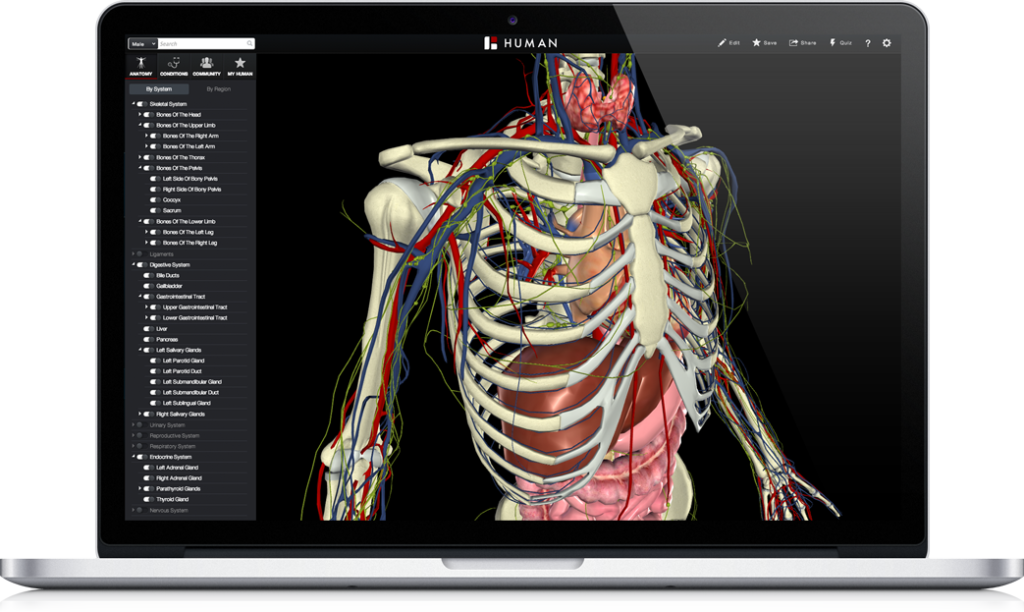
In between modeling the actual and modeling the conceptual lies an intermediary: 3D visualization softwares of anatomy(BioDigital) or of biological concepts (BioRender, BIOZONE) and more. These virtual platforms offer interactive ways of learning about the human anatomy and biological processes that occur within it. These programs are a lot more inventive and creative than simple replications of objects where the model itself only exists as a copy of the information. BioDigital, for instance, not only accurately models human anatomy, it can also narrate a particular illness that happens within the human anatomy and its condition by using indicators and different graphic elements that facilitate comprehension of the information. Graphics and design become fundamental aspects in helping engage the viewer with understanding the process and location of an illness or distinguishing different components, particles, or systems that are a part of the illness, and etc. Rendering and textural choices also serve as a certain aesthetic choice in finalizing the model that could distinguish their style from other visualization softwares.
Modeling the imaginary or conceptual is often used when the question is no longer how to replicate the formal structures of the object but rather experimental: how to model and calculate cell interaction, tissue growth, fracture healing. These questions are then posed into a hypothesis and generated into the model, initiating a virtual experiment. G. Wayne Brodland’s article, “How Computational Models Can Help Unlock Biological Systems” presents information on how digital modeling aids cell biology, especially on learning how cells interact with one another. After the invention of computers, different types of models were designed to visualize cell interaction in different ways. Cellular Potts Model, Finite Element Method, Monte Carlo models are examples of different systems of modeling that could provide different solutions to the experiment.
However, before the model could be generated, the researcher or scientist needs to choose specific “state variables”(what I thought of as an artist’s decision, choice in fabrication, composition), as the article mentions, that could then be calculated into computational codes. These “state variables”, or experimenter’s choice of which elements to keep and which to take out. These include, “surface points that defined the exterior form of the embryo, gene expression patterns, cytoskeletal morphologies, and cell fabric (size, shape and directionality) information”[3]. Data that are collected from these variables are plugged into a computational model of one’s choice that can most effectively bring expected results.
One of these models that are used in modeling for engineering, physics, as well as cell biology is the Finite Element Method (FEM). Department of Dental Sciences and Surgery and Faculty of Medicine and Surgery at the University of Bari discusses the Finite Element Method and its uses in Mechanobiology, science incorporating biology, engineering, and physics. This particular model can provide physical and structural information of the subject as well as calculate any changes that are applied onto the form. Individual geometrical points in FEM are defined as ‘nodes’ and the overall geometry is made up of tetrahedra that consists of multiple nodes that ultimately form a finite element mesh. After the modeling stage, the model is varnished and edited with computer graphics to further finalize the texture and finish of the model. The same information, such as cell interaction patterns can be visualized in different ways using the Cellular Potts Model which models in a lattice structure or Monte Carlo Models that models with experimental or random measurements. An example below shows cell deformation when a force is being applied onto the structure using the Finite Element Method.
By looking at various digital models that help visualize conceptual ideas in biology, we notice scientists along with artists expanding their imagination in creating different ways of virtual experimentation. Experiments that are generated in digital spaces allow for infinite runs as well as infinite changes, various modeling types, and rendering or graphic finish of one’s choice. The end result is a creation of a model that is unique, simplified, and altered from the original, thus inventing a new representation of the original form, idea or concept.
[1] Barazzetti, Luigi, Marco Gianinetto, and Marco Scaioni. “Automatic Image-Based 3D Modeling for Medical Applications.” 2012 5th International Conference on BioMedical Engineering and Informatics, 2012. https://doi.org/10.1109/bmei.2012.6512942.
[2] Barazzetti, Luigi, Marco Gianinetto, and Marco Scaioni. “Automatic Image-Based 3D Modeling for Medical Applications.” 2012 5th International Conference on BioMedical Engineering and Informatics, 2012. https://doi.org/10.1109/bmei.2012.6512942.
[3] Brodland, G. Wayne. “How Computational Models Can Help Unlock Biological Systems.” Seminars in Cell & Developmental Biology 47-48 (2015): 62–73. https://doi.org/10.1016/j.semcdb.2015.07.001.
[4] 2018, BioDigital. “BioDigital Human: Anatomy and Health Conditions in Interactive 3D.” BioDigital. Accessed March 28, 2020. https://www.biodigital.com/product.
[5] “Overview.” Finite Element Modelling of A Living Cell. Accessed April 4, 2020. http://s1148054.weebly.com/overview.html.