Introduction:
3D printing has revolutionized many industries, due to its ability to quickly prototype complex objects. One of the most exciting and rapidly developing applications of 3D printing is in the field of biotechnology, specifically in the creation of 3D-printed tissue. This innovative technology is helping to bridge the gap between engineering and health, offering potential solutions for organ transplant shortages, medical testing, and personalized medicine.
What is 3D Printed Tissue?
3D printing is a process where material is deposited layer by layer to create a three-dimensional object. Traditionally, 3D printing has been used for producing physical prototypes and parts in fields like engineering and manufacturing. Most commonly, 3D printing uses plastic filament, clay, or resin to print objects. However, in the context of tissue printing, the process is adapted to create living cells and biological materials. Instead of printing plastic or metal, specialized 3D printers use bioinks, a combination of living cells, growth factors, and other biomaterials.
The goal of 3D printing tissue, sometimes referred to as bioprinting, is to create functional biological structures that mimic the complexity of natural tissue. These tissues can be used for various purposes, such as testing new drugs, creating organ models for surgery, or even replacing damaged or diseased tissues in the human body. Unlike traditional methods of growing tissues in a lab, 3D printing allows for precise control over the shape, size, and arrangement of the cells, enabling the creation of more complex and realistic tissues. The vascular innovations in 3D printing most importantly allow tissue to grow bigger and live for longer. In lab grown tissue as it grows thicker, it becomes increasingly difficult to properly disperse oxygen and nutrients to feed the cells. Harvard bioengineers Jennifer Lewis and Christopher Chen further emphasize this saying, “Solving this problem could dramatically speed up the development of implantable human tissues that can save lives.”
The Process:
1. Designing the Model: Researchers begin by creating a digital model of the tissue they want to print. This is done using 3D modeling software, where they map out the desired structure, including the arrangement of different cell types and the vascular system (blood vessels) that will supply nutrients to the tissue.
2. Preparing the Bioink: Bioinks are made from a mixture of cells and biomaterials called hydrogels. Hydrogels can be a range of materials like collagen, gelatin or alginate, all of which act as scaffolds to support the cells as they grow and develop. The bioink must be carefully formulated to ensure that the cells can survive and reproduce in the printed structure. Depending on the printing process, the biofabricated object will be treated with an ionic solution or UV light to encourage crosslinking (the process in which the hydrogel polymers connect and solidify).
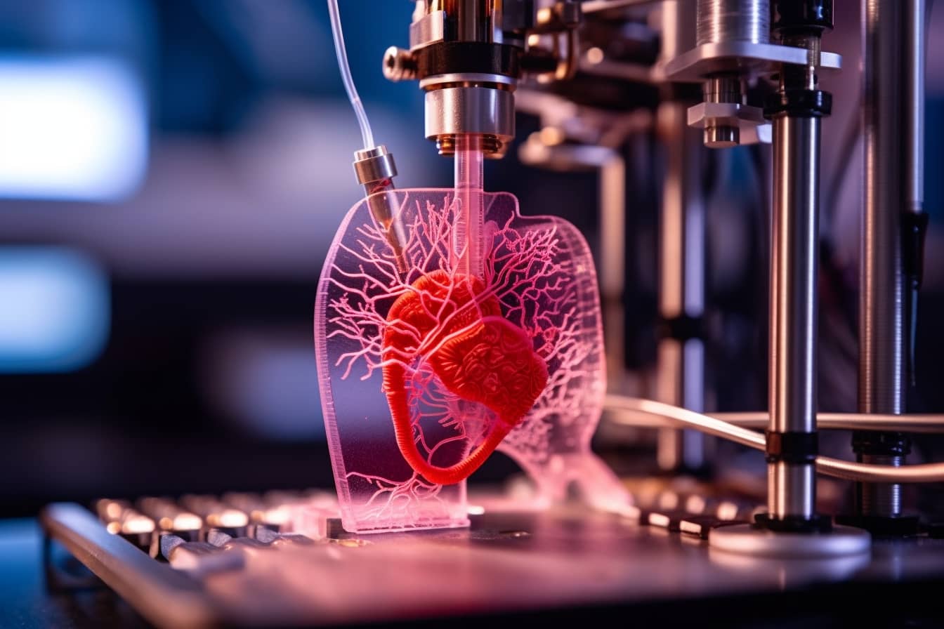
3. Printing the Tissue: Using a specialized 3D printer or bioprinter, the bioink is deposited layer by layer, according to the digital model. The printer uses different techniques to create the structure, such as inkjet-based, extrusion-based, or laser-assisted methods. Once printed, the tissue is placed in a bioreactor, a controlled environment that provides nutrients and oxygen to encourage cell growth and maturation.
4. Maturation and Growth: After printing, the tissue needs time to mature. This is where the cells begin to grow, organize, and form functional tissue. Researchers may also introduce growth factors or additional support structures to help guide the tissue toward its intended function.
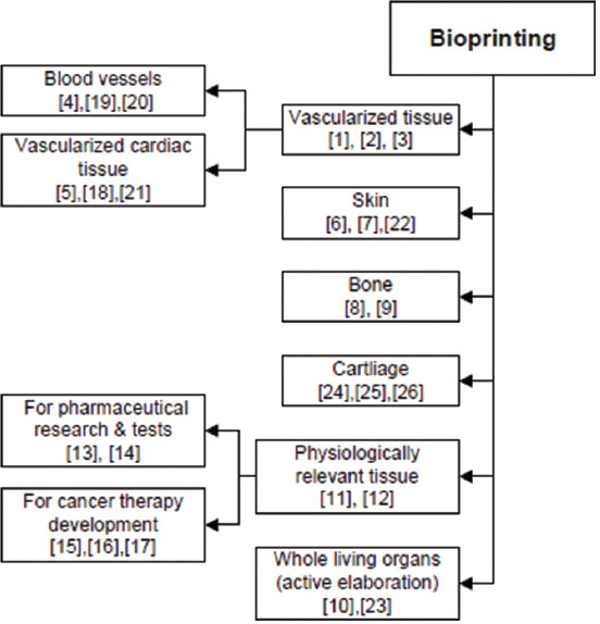
Bioprinting Applications
1. Drug Testing and Development: One of the earliest applications of 3D-printed tissue is in drug testing. Traditional methods of testing new pharmaceuticals often involve animal models or two-dimensional cell cultures. However, these models don’t fully replicate the complexities of human tissue. 3D-printed tissues, on the other hand, can mimic the real environment of human cells more closely, providing researchers with better data on how a drug will perform in the human body. This approach also reduces the need for animal testing and could lead to more effective and safer drugs.
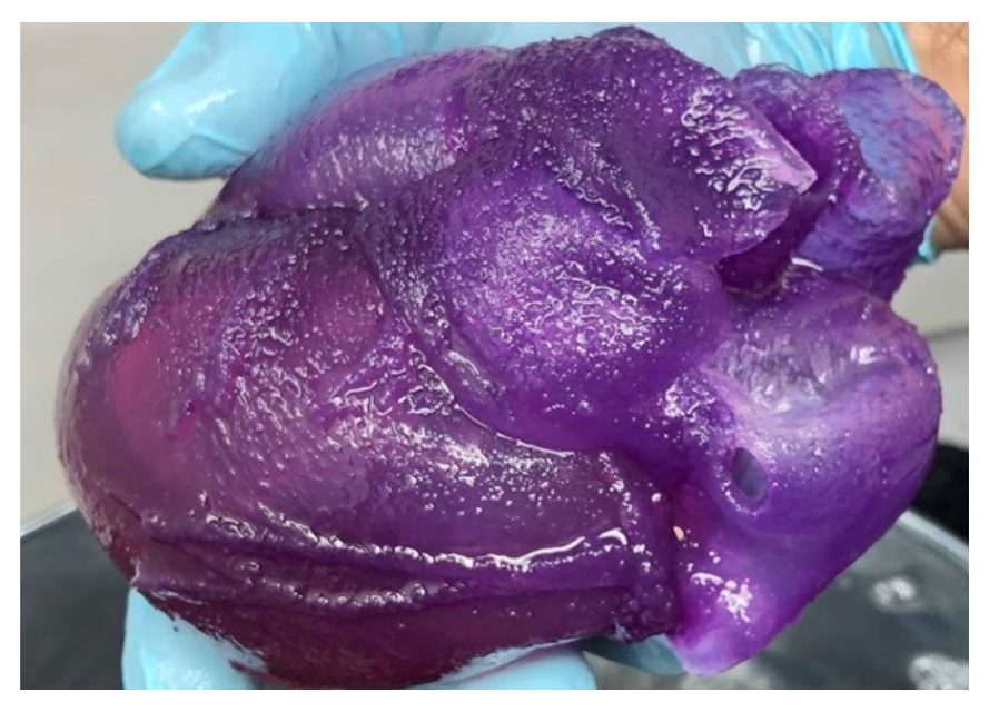
2. Organ and Tissue Replacement: One of the most ambitious goals of 3D tissue printing is the ability to print entire organs, such as kidneys, livers, or hearts that could be transplanted into patients. While printing fully functional organs is still far from reality, researchers are making significant progress in printing smaller tissues, such as skin, cartilage, and liver tissue. In the future, this could help address the global shortage of organ donors and allow for more personalized medicine, where organs are created to match the recipient’s genetic makeup.
3. Surgical Planning and Education: 3D printing also has applications in **medical training and surgical planning**. By printing detailed models of organs or tissues based on a patient’s medical scans, doctors can practice complex surgeries before performing them on a real patient. This allows for more precise and less invasive surgeries, improving patient outcomes. Surgeons can also use these models to visualize and plan surgeries in a way that was never possible before.
4. Skin Grafting and Wound Healing: 3D printing has shown promise in regenerating skin tissue for patients with severe burns or wounds. In 2018, researchers successfully used a 3D printer to create a layer of living skin cells, which could be grafted onto burn victims. This skin was printed with an embedded network of blood vessels, allowing it to integrate better with the body’s existing tissue. This method could significantly speed up the recovery process for patients and reduce the risk of infection or rejection.
5. Personalized Medicine: 3D printing enables personalized treatment by creating tissues that match a patient’s unique genetic profile. For example, a patient’s tumor cells can be printed to create a model, allowing doctors to test different treatments to see which one works best for that individual. This approach could revolutionize cancer treatment, providing more targeted and effective therapies.
Conclusion
The 3D printing of tissue is an exciting and rapidly advancing field with the potential to transform medicine, from drug testing to organ transplantation. While there are still many challenges to overcome, the applications of this technology could greatly improve patient care, reduce the reliance on animal testing, and provide personalized medical treatments. As 3D printing technology continues to evolve, it could potentially revolutionize how we approach healthcare and biotechnology, offering new solutions to some of the most pressing challenges in modern medicine.
Sources:
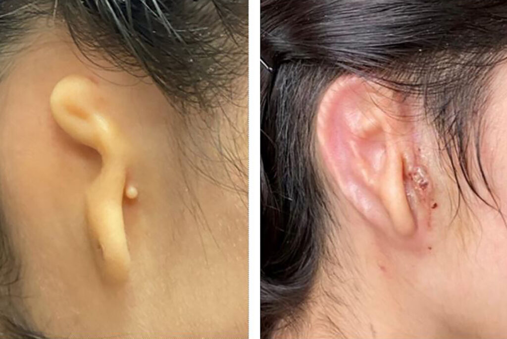
https://www.nytimes.com/2022/06/02/health/ear-transplant-3d-printer.html%20
https://engineering.cmu.edu/news-events/news/2020/11/18-3d-printed-heart.html
https://www.sciencedirect.com/science/article/pii/S2590183424000127
https://www.sciencedirect.com/science/article/pii/S2666964123000206
1 thought on “Building Life: The Future of 3D Printed Tissues”
Comments are closed.